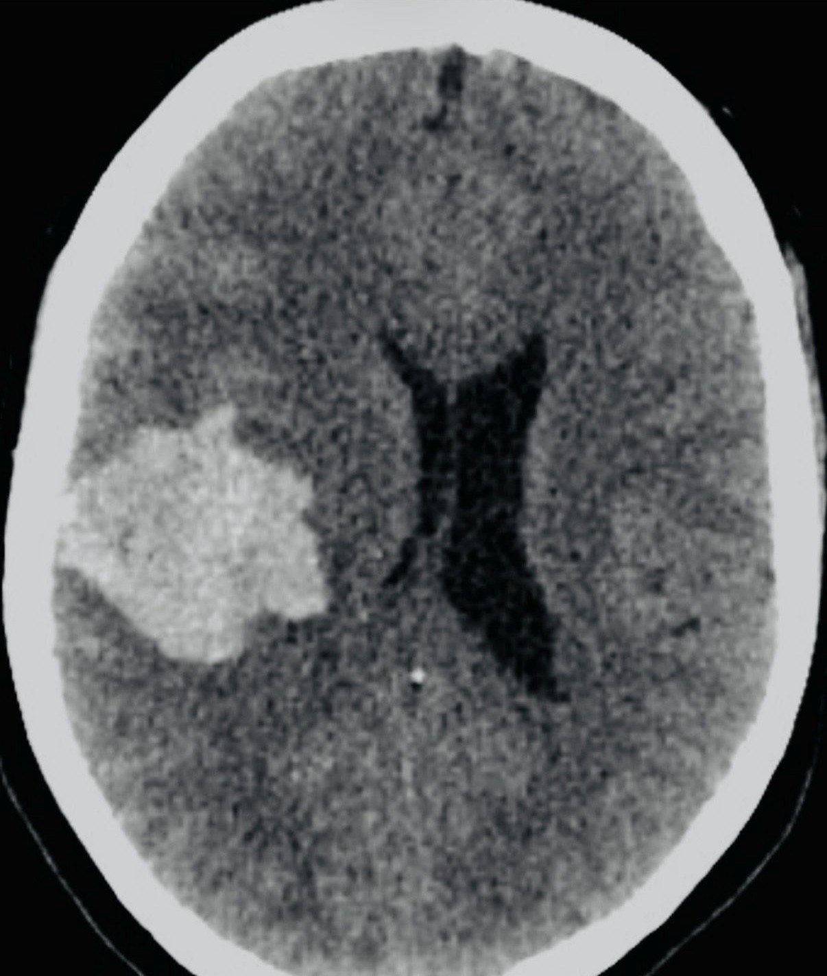
means intracranial arteries: rupture warning is often sudden worst headache, collapse or stroke-like symptoms.
Aneurysm detection and next steps
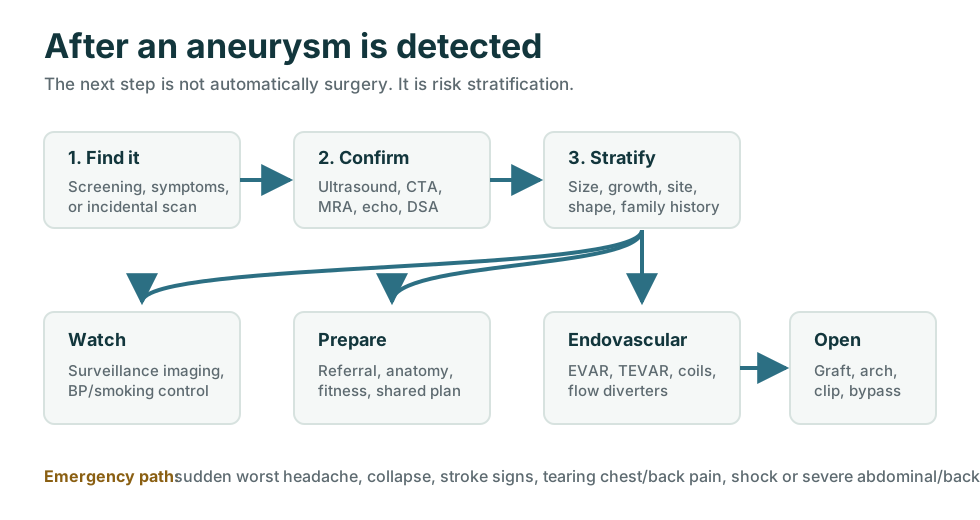
First choose the aneurysm pathway: intracranial/brain or aortic.
A brain aneurysm, an abdominal aortic aneurysm, a thoracic aortic aneurysm, a leak/rupture and an aortic dissection are different scenarios. Blood flow is the body's oxygen supply; when a vessel tears, leaks, ruptures or blocks a branch, the brain, heart, kidneys, bowel or legs can lose oxygen fast. That can mean life or death, brain damage, heart ischemia, organ failure or limb loss.
Choose brain or aorta
Incidental scan finding
Emergency signs
Aortic dissection
Leak or rupture
Survival rates
Operate or observe
Repair thresholds
IntracranialAneurysm.com
Intracranial / brain
Head, brain arteries, subarachnoid hemorrhage risk
Often CTA, MRA or catheter angiography; decisions may involve neurosurgery or interventional neuroradiology.
Aortic: abdominal / thoracic
Abdominal aortic, thoracic, arch, root or iliac aneurysm
AAA often starts with ultrasound; thoracic, arch and root aneurysms often need CTA, MRA or echocardiography. Decisions may involve vascular or cardiac aortic teams.
means abdominal aortic aneurysm: ultrasound is common for screening and surveillance.
means thoracic aortic aneurysm: CTA, MRA and echo often define chest/root anatomy.
means leak, rupture, dissection, new severe pain or organ compromise: emergency beats elective thresholds.
Aortic and body aneurysms
Aortic aneurysms are managed by location, size, growth and symptoms.
This pathway covers abdominal aortic aneurysm, thoracic aortic aneurysm, aortic root, ascending aorta, arch, descending thoracic, thoracoabdominal and iliac aneurysms. A small incidental aneurysm can be safer to monitor than to operate on. A symptomatic, rapidly enlarging, ruptured or above-threshold aneurysm is different.
Aortic locations to name clearly
The aortic/body questions
Where is it? What is the maximum diameter? Is it truly an aneurysm or an ectatic vessel? Is it growing? Is it causing symptoms? Is there connective tissue disease, bicuspid aortic valve, family history or infection? Is the anatomy suitable for endovascular repair?
Emergency first
Some aneurysm symptoms are not watch-and-wait problems.
Rupture or dissection can kill quickly. Do not drive yourself to clinic with these symptoms.
Aortic dissection
Dissection is a tear in the aortic wall, not just a large aneurysm.
An aneurysm is a dilated vessel. A dissection is blood tracking through a tear in the aortic wall. They can overlap, but they are not the same problem. Acute dissection is usually handled as an emergency until proven otherwise.
Leak, rupture, elective or emergency
The same aneurysm can be a planned repair problem or an emergency problem.
Elective aneurysm care is about preventing future rupture when the risk of waiting becomes higher than the risk of repair. Emergency care is different: leak, rupture, dissection, shock, uncontrolled pain or organ compromise can override usual size thresholds.
Survival rates and risk context
Numbers change sharply between elective repair and emergency rupture or dissection.
These figures are broad published ranges, not a personal prediction. Age, frailty, comorbidities, pre-existing dementia, prior strokes, baseline independence, shock, kidney function, lung disease, heart disease, diabetes, anticoagulants, anatomy, hospital volume, repair type and how quickly treatment starts can change the odds a lot.
Often low single-digit early mortality in selected patients
Modern elective abdominal aortic aneurysm repair is much safer than rupture repair. Published estimates often put 30-day mortality around 1% to 2% for lower-risk EVAR/open patients, but substantially higher for high-risk open repair.
Overall mortality is often quoted around 80% to 90%
Many patients with ruptured abdominal aortic aneurysm die before reaching hospital. Those who reach a vascular center may survive emergency EVAR or open repair, but shock and organ failure drive risk.
Minutes and hours matter
Untreated acute type A dissection has historically been described as rising roughly 1% to 2% mortality per hour early on. Even with surgery, it remains a high-risk emergency.
Uncomplicated and complicated are different
Uncomplicated type B dissection may start with intensive medical therapy. Rupture, malperfusion, uncontrolled pain, expansion or high-risk anatomy can move care toward TEVAR or surgery.
Center and anatomy matter
Thoracic, arch and thoracoabdominal operations vary widely. Root/ascending open surgery, descending TEVAR, open thoracoabdominal repair and hybrid arch repair do not have one single survival number.
Ruptured and unruptured are separate worlds
An unruptured incidental intracranial aneurysm may be watched or treated electively. Rupture with subarachnoid hemorrhage is a neurologic emergency with very different survival and disability risks.
Risk of not operating vs risk of intervention
The real question is not “operate or do nothing”; it is “which risk is bigger now?”
Observation is an active plan when the aneurysm is small, stable and asymptomatic. Intervention becomes more attractive when the chance of rupture, dissection, leak, embolism, compression or organ compromise starts to outweigh the risk of the operation itself.
Follow-up ultrasound, CT or MRI
Surveillance means choosing the right scan at the right interval.
The follow-up test depends on aneurysm location, size, growth, repair history, kidney function, contrast risk, radiation exposure and whether the question is simple diameter, branch-vessel anatomy, leak, endoleak or dissection change.
Ultrasound
Often the first follow-up test for small abdominal aortic aneurysms. It is quick, noninvasive and avoids radiation, but may not answer complex anatomy questions.
CT angiogram
Used when symptoms, rapid growth, rupture concern, preoperative planning, iliac involvement or device planning requires detailed anatomy.
CTA, MRA or echo
Thoracic aneurysms often need CT or MRI for the full aorta. Echocardiography is useful for the aortic root, ascending aorta and valve-related disease.
Endoleak surveillance
After stent-graft repair, follow-up looks for endoleak, graft migration, sac growth, branch issues and device integrity. CT, ultrasound or contrast-enhanced ultrasound may be used depending on the case.
CT or MRI schedule
Dissection follow-up watches the false lumen, branch vessels and later aneurysmal enlargement. CT or MRI is commonly repeated after the acute phase and then periodically if stable.
MRA, CTA or DSA
Intracranial aneurysm follow-up usually uses MRA or CTA when observing, with catheter angiography when the anatomy or treatment result needs higher detail.
High-risk decisions and support
Aneurysm, rupture and dissection decisions are high-mortality, high-consequence problems.
Emergency repair, ICU care, anesthesia, neurologic outcome, long-term disability, family communication and advance-care preferences may all matter. This is critical infrastructure for a human body: when the main oxygen supply fails, the outcome can change minute by minute. Survival is not the only outcome; brain injury, paralysis, kidney failure, bowel ischemia, limb loss and loss of independence can matter just as much.
AnonamedBroader medical navigation and related health-site network.
AdvanceCareDirective.comPlanning preferences before high-risk surgery, ICU care, dementia/stroke-related loss of capacity or catastrophic deterioration.
AnesthesiaRisks.comRisk context for major vascular, cardiac, neurosurgical and emergency operations.
TheRisks.comGeneral risk framing for rare-but-severe outcomes, uncertainty, consent and tradeoff decisions.
TheVascularSurgeons.comVascular specialist pathway for abdominal, iliac, descending thoracic and peripheral aneurysm problems.
TheNeurosurgeons.comBrain aneurysm, subarachnoid hemorrhage, clipping, coiling and neurosurgical decision support.
TheHospitals.orgHospital-level context for emergencies, imaging, operating rooms, ICU and transfer decisions.
Confirm and measure
The next test depends first on whether it is intracranial/brain, abdominal aortic, or thoracic/chest aortic.
Aneurysm care begins by confirming the location with the right imaging and measuring the maximum diameter consistently. If leak, rupture or dissection is suspected, the scan is also looking for bleeding, branch-vessel compromise and emergency anatomy.
Duplex ultrasound
Common for AAA screening and surveillance. It is noninvasive and avoids radiation, but bowel gas or body habitus can limit views.
CT angiogram
Maps diameter, neck length, branch vessels, iliac access and leak/rupture concerns. Often the key preoperative planning test.
MRI / MRA
Useful when radiation or iodinated contrast is a concern, and for some thoracic and brain vessel assessment.
Echocardiography
Assesses aortic valve, root and ascending aorta, especially with bicuspid valve or Marfan-like syndromes.
Acute CTA pathway
When dissection is suspected, imaging is about locating the tear, branch-vessel compromise and rupture risk, not just measuring aneurysm diameter.
CTA, MRA, DSA
CTA and MRA can detect many intracranial aneurysms. Catheter angiography gives high-detail anatomy when noninvasive tests are not enough.
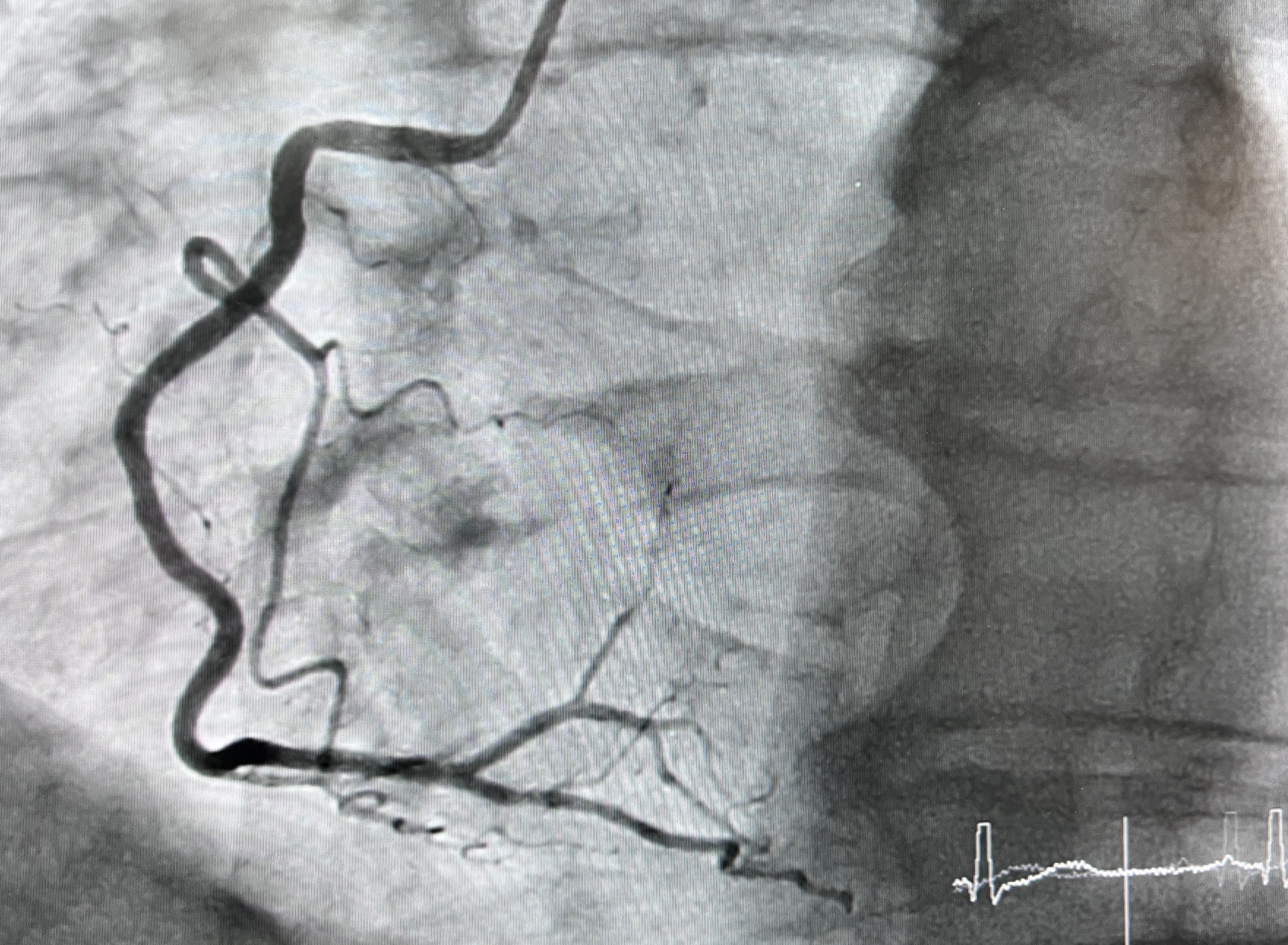
Aortogram / angiogram
Catheter angiography is less often a first screening test, but may be used during planning or treatment.
Whole-body MRI and incidental findings
Incidental aneurysm findings still need to be sorted into brain or aortic/body pathways.
Whole-body MRI, cancer-screening MRI, CT calcium scoring, spine scans and abdominal imaging can unexpectedly report a possible aneurysm. That can be useful, but the finding usually needs targeted confirmation before anyone decides on surveillance intervals or repair.
Do not jump from incidental finding to operation
A commercial whole-body MRI can create a useful warning, but it can also overcall or under-measure vascular disease. Aneurysm decisions need dedicated imaging, prior-scan comparison, risk factors and specialist review.
The American College of Radiology does not recommend total-body screening MRI for people without symptoms, risk factors or family history because the evidence of benefit is insufficient and non-specific findings can lead to extra testing and expense.
Watch and wait
Surveillance is active management, not doing nothing.
Small aneurysms are often monitored because repair itself has risk. The plan usually includes imaging intervals, blood pressure control, smoking cessation, statin/vascular risk management when appropriate, and instructions about emergency symptoms.
| Finding | Typical next step | Why it matters |
|---|---|---|
| AAA 3.0 to 3.9 cm | Ultrasound surveillance, often about every 3 years. | Rupture risk is usually low, but growth must be tracked. |
| AAA 4.0 to 4.9 cm in men, 4.0 to 4.4 cm in women | Often annual ultrasound. | Growth risk rises as diameter increases. |
| AAA at least 5.0 cm in men or 4.5 cm in women | Often imaging about every 6 months and vascular review. | Approaches common repair thresholds. |
| Thoracic aneurysm below threshold | Repeat imaging schedule based on site, size, cause and growth. | Root, ascending, arch and descending aneurysms behave differently. |
| Small unruptured brain aneurysm | Neurosurgery/interventional neuroradiology review, risk scoring and interval MRA/CTA when observation is chosen. | Rupture risk depends on size, shape, site, patient age, smoking, blood pressure and family history. |
Criteria for intervention
Repair is considered when rupture/dissection risk outweighs treatment risk.
Exact thresholds vary by sex, body size, anatomy, inherited syndrome, valve disease, symptoms, surgical center experience and national guidelines. These are practical guideposts, not personal orders.
Endovascular or open
The operation is chosen for anatomy as much as for size.
Endovascular repair is less invasive but needs suitable landing zones and lifelong surveillance. Open repair is more invasive but can be more durable or necessary for anatomy that endovascular devices cannot safely seal.
EVAR
A stent graft is placed through groin arteries to exclude the aneurysm sac. Good anatomy matters: neck length, angulation, iliac access and branch vessels.
TEVAR
Endovascular stent grafting for selected descending thoracic aneurysms. Arch or visceral branch involvement may require complex/hybrid approaches.
Open repair or TEVAR
Type A dissection commonly means urgent open cardiac/aortic surgery. Complicated type B dissection may be treated with TEVAR when anatomy and timing fit.
Open aortic repair
The diseased aorta is replaced with a graft. It may be required for ascending aorta, arch, infected aneurysm, unsuitable anatomy or younger/complex patients.
Coiling, flow diversion, clipping
Endovascular and microsurgical choices depend on aneurysm neck, branch vessels, rupture status, age and center expertise.
Intracranial and brain aneurysms
If the report says intracranial, brain or cerebral aneurysm, use the brain pathway.
Brain aneurysm decisions are different from aortic aneurysm decisions. The key issues are rupture history, subarachnoid hemorrhage symptoms, artery location, neck shape, branch vessels, CTA/MRA/DSA anatomy, and whether observation, coiling, flow diversion or clipping is safest. For the deeper brain-specific guide, use IntracranialAneurysm.com.
Sources
Guidelines and patient resources used for this guide.
2022 ACC/AHA Aortic Disease GuidelineDiagnosis, surveillance, thresholds and repair principles for aortic disease.
ACC aortic dissection key pointsType A, type B, acute aortic syndromes and follow-up imaging principles.
JAMA Network Open elective AAA repair riskRisk-stratified 30-day mortality estimates for elective AAA repair.
IRAD type A dissection mortalityEarly mortality patterns and urgency in acute type A aortic dissection.
USPSTF AAA screeningWho benefits from abdominal aortic aneurysm screening and why ultrasound is used.
ACC key perspectivesPlain-language summary of aortic disease guideline highlights.
MedlinePlus brain aneurysmSymptoms, CTA/MRA/angiography and treatment overview.
NINDS cerebral aneurysmsPatient information on cerebral aneurysm diagnosis and treatment.
AAFP ruptured intracranial aneurysm reviewRecognition, evaluation and common SAH grading systems including Hunt and Hess and modified Fisher.
ACR total-body MRI statementEvidence and incidental-finding concerns in asymptomatic screening.
TheVascularSurgeons.comFind vascular specialists and aortic teams.